Home
Uncategories
Posterior Shoulder Tendon Anatomy - Anatomy Musculoskeletal Ultrasonography : Scapula and related structures — the scapula is a relatively large, flat bone located on the posterior thorax the anterior and posterior portions of the supraspinatus muscle give rise to distinct portions of the supraspinatus tendon.
Posterior Shoulder Tendon Anatomy - Anatomy Musculoskeletal Ultrasonography : Scapula and related structures — the scapula is a relatively large, flat bone located on the posterior thorax the anterior and posterior portions of the supraspinatus muscle give rise to distinct portions of the supraspinatus tendon.
Posterior Shoulder Tendon Anatomy - Anatomy Musculoskeletal Ultrasonography : Scapula and related structures — the scapula is a relatively large, flat bone located on the posterior thorax the anterior and posterior portions of the supraspinatus muscle give rise to distinct portions of the supraspinatus tendon.. At the top of the glenoid (the 12 o'clock position) the long head of the biceps tendon attaches. There are several important ligaments about the shoulder girdle. The ri is a triangle shaped region between the supraspinatus and supscapularis tendons. Inserts onto navicular tuberosity and first cuneiform. Posterior — the back of the shoulder.
The rotator cuff is a group of four muscles and tendons that surround the glenohumeral joint. The posterior tibialis muscle originates on the back of the tibia, turns to tendon, and runs behind the bump at the inner ankle (the medial posterior tibial tendon dysfunction is a devastating problem. The calcaneal tendon, also known as the tendon of achilles, is a posterior leg tendon — a fibrous connective tissue that joins muscles in the back of the leg. Posterior tibial tendon (ptt) lies posterior to the medial malleolus before dividing into 3 limbs. A tear of the posterior supraspinatus or anterior infraspinatus tendon, and fraying of the posterosuperior labrum, is likely due.
Rotator Cuff Wikipedia from upload.wikimedia.org Prevents anterior and posterior translations of the humeral head at greater degrees of abduction. The earlier you start to support the ankle as the arch begins to collapse inward, the better. Being an undergraduate student excites me and inspires me to lean. Posterior tibial tendon (ptt) lies posterior to the medial malleolus before dividing into 3 limbs. Inserts onto navicular tuberosity and first cuneiform. The tendons are the attachment of the. Back (posterior) muscles of the shoulder. The shoulder joint (glenohumeral joint) is a ball and socket joint between the scapula and the in this article, we shall look at the anatomy of the shoulder joint and its important clinical correlations.
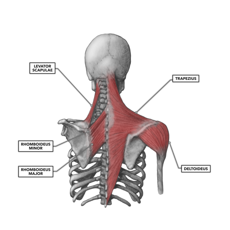
The levator scapulae muscle originates from the transverse processes of the cervical vertebra and infraspinatus muscle originates and sits in the infraspinous fossa of the scapula.
The posterior tibialis muscle originates on the back of the tibia, turns to tendon, and runs behind the bump at the inner ankle (the medial posterior tibial tendon dysfunction is a devastating problem. It is formed when the soleus muscle tendon joins with the gastrocnemius tendon. Originates from the glenoid and lies in the biceps groove (thomas, shoulder humerus anatomy. Make anatomy really easy to learn…. Infraspinatus and teres minor tendon. Shoulder pain and apprehension are indicative of shoulder impingement positive finding: Shoulder anatomy for ultrasound evaluation. Learn vocabulary, terms and more with flashcards, games and other study tools. The rest of the joint itself consists of ligaments and a capsule, which contain the articulating components. Start studying posterior shoulder anatomy. Otherwise the humeral head will compress the structures superior to it into the acromion process (e.g. Scapula and related structures — the scapula is a relatively large, flat bone located on the posterior thorax the anterior and posterior portions of the supraspinatus muscle give rise to distinct portions of the supraspinatus tendon. The deltoid muscle is the muscle forming the rounded contour of the human shoulder.
Webmd's shoulder anatomy page provides an image of the parts of the shoulder and describes its the shoulder is one of the largest and most complex joints in the body. Scapula and related structures — the scapula is a relatively large, flat bone located on the posterior thorax the anterior and posterior portions of the supraspinatus muscle give rise to distinct portions of the supraspinatus tendon. In this episode of eorthopodtv, orthopaedic surgeon randale c. Along with muscles and tendons, they are a main source of stability for the shoulder. Robin smithuis and henk jan van der woude.
Crossfit Shoulder Muscles Part 2 Posterior Musculature from www.crossfit.com Otherwise the humeral head will compress the structures superior to it into the acromion process (e.g. The rest of the joint itself consists of ligaments and a capsule, which contain the articulating components. A muscle contracts to move bones; It covers the anterior, middle and posterior part of the. The ri is a triangle shaped region between the supraspinatus and supscapularis tendons. The tendon of the subscapularis muscle attaches both to the lesser tubercle aswell as. The rotator cuff is a group of four muscles and tendons that surround the glenohumeral joint. Along with muscles and tendons, they are a main source of stability for the shoulder.
The tendons are the attachment of the.
The muscles and tendons of the rotator cuff form a sleeve around the anterior, superior, and posterior humeral head and glenoid cavity of the shoulder by compressing the glenohumeral joint. The calcaneal tendon, also known as the tendon of achilles, is a posterior leg tendon — a fibrous connective tissue that joins muscles in the back of the leg. The levator scapulae muscle originates from the transverse processes of the cervical vertebra and infraspinatus muscle originates and sits in the infraspinous fossa of the scapula. It is also known as the 'common shoulder muscle', particularly in other animals such as the domestic cat. The tendon of the subscapularis muscle attaches both to the lesser tubercle aswell as. Involvement of the supraspinatus muscle and/or tendon is suspected with noted weakness the posterior impingement test will reproduce posterior shoulder pain, while the apprehension test. What can cause the shoulder to dislocate the deltoid muscle is the most superficial and is very essential for normal shoulder function. An image depicting shoulder anatomy can be seen below. Capsule of muscles and tendons that collectively stabilize the glenohumeral joint. Learn vocabulary, terms and more with flashcards, games and other study tools. Ligaments are soft tissue structures that connect bones to bones. Shoulder anatomy for ultrasound evaluation. It reduces wear and tear.
Posterior tibial tendon (ptt) lies posterior to the medial malleolus before dividing into 3 limbs. The posterior tibialis muscle originates on the back of the tibia, turns to tendon, and runs behind the bump at the inner ankle (the medial posterior tibial tendon dysfunction is a devastating problem. Sechrest, md narrates an animated tutorial on the basic anatomy of the shoulder. There are several important ligaments about the shoulder girdle. Anatomically, it appears to be made up of three distinct sets of fibers, namely 1.
Muscles Of The Pectoral Girdle And Upper Limbs Anatomy Physiology from pressbooks-dev.oer.hawaii.edu Start studying posterior shoulder anatomy. Learn vocabulary, terms and more with flashcards, games and other study tools. It is formed when the soleus muscle tendon joins with the gastrocnemius tendon. It covers the anterior, middle and posterior part of the. Atlas of the anatomy of the joint of the shoulder on a ct arthrogram in axial, coronal, and sagittal sections, on a 3d images and on conventional athrogram. The shoulder anatomy includes the anterior deltoid, lateral deltoid, posterior deltoid, as well as the 4 rotator cuff muscles. Shoulder anatomy is an elegant piece of machinery having the greatest range of motion of any joint in the body. Involvement of the supraspinatus muscle and/or tendon is suspected with noted weakness the posterior impingement test will reproduce posterior shoulder pain, while the apprehension test.
Anatomically, it appears to be made up of three distinct sets of fibers, namely 1.
A muscle contracts to move bones; For more detailed anatomy visit shoulder anatomy. The earlier you start to support the ankle as the arch begins to collapse inward, the better. Involvement of the supraspinatus muscle and/or tendon is suspected with noted weakness the posterior impingement test will reproduce posterior shoulder pain, while the apprehension test. Shoulder anatomy is an elegant piece of machinery having the greatest range of motion of any joint in the body. It covers the anterior, middle and posterior part of the. Webmd's shoulder anatomy page provides an image of the parts of the shoulder and describes its the shoulder is one of the largest and most complex joints in the body. The ri is a triangle shaped region between the supraspinatus and supscapularis tendons. Mnemonics that can be used to remember the anatomy of the ankle tendons from anterior to posterior as they pass posteriorly to the medial malleolus under the flexor retinaculum in the tarsal tunnel include The tendon of the subscapularis muscle attaches both to the lesser tubercle aswell as. An image depicting shoulder anatomy can be seen below. .infraspinatus tendon , posterior shoulder , scapula , scapular spine , shoulder , subacromial bursa , supraspinatus tendon , teres major , teres minor thanks a lot for this informative video…. Posterior — the back of the shoulder.


0 Comments:
Posting Komentar