Home
Uncategories
Posterior Rib Cage Muscles - BIO 141 Muscles - Biology 141 with Rodgers at Northern ... / Measuring rib cage and abdominal movement is the most common technique for assessing thoracic cage and pulmonary mechanics.
Posterior Rib Cage Muscles - BIO 141 Muscles - Biology 141 with Rodgers at Northern ... / Measuring rib cage and abdominal movement is the most common technique for assessing thoracic cage and pulmonary mechanics.
Posterior Rib Cage Muscles - BIO 141 Muscles - Biology 141 with Rodgers at Northern ... / Measuring rib cage and abdominal movement is the most common technique for assessing thoracic cage and pulmonary mechanics.. Perspective front and back views. The chest muscles are specifically modified due to modifications in axial. Intercostal muscles are a group of muscles which exist in the intercostal space and help create from lateral border of sternum to the angle of rib (posteriorly it continues as posterior. The pain may occur immediately upon injury or. It is the area of articulation with the transverse process of the vertebra.
Did you know the rib cage plays a role in posture alignment? For this purpose isolated strips of rib cage elevator muscle of variations in the musculoskeletal system exist in different classes of animal. The secondary muscles of respiration are the anterior/medial scalenes, serratus posterior the thoracic spine and rib cage: Central slip and mallet finger management online course: The posterior or back muscles perform a wide range of functions, including movement of the shoulder, head, and neck and assisting in respiration, posture.
8. Muscles of the Spine and Rib Cage | Musculoskeletal Key from musculoskeletalkey.com Review the anatomical characteristics of the rib and ribcage in this interactive tutorial and test your knowledge in the quiz. The rib cage is composed by sternum, costal cartilages, and ribs connected to the thoracic vertebrae. The posterior or back muscles perform a wide range of functions, including movement of the shoulder, head, and neck and assisting in respiration, posture. We're going to look at a pair of them that do just that: Did you know the rib cage plays a role in posture alignment? Human 3/4 body skeleton with muscles, veins and arteries. It is supplied with blood by the lowest posterior intercostal artery, the subcostal artery articular cartilage of left superior articular facet of sacrum. These spaces are filled by intercostal muscles, and they also contain intercostal nerves and blood vessels.
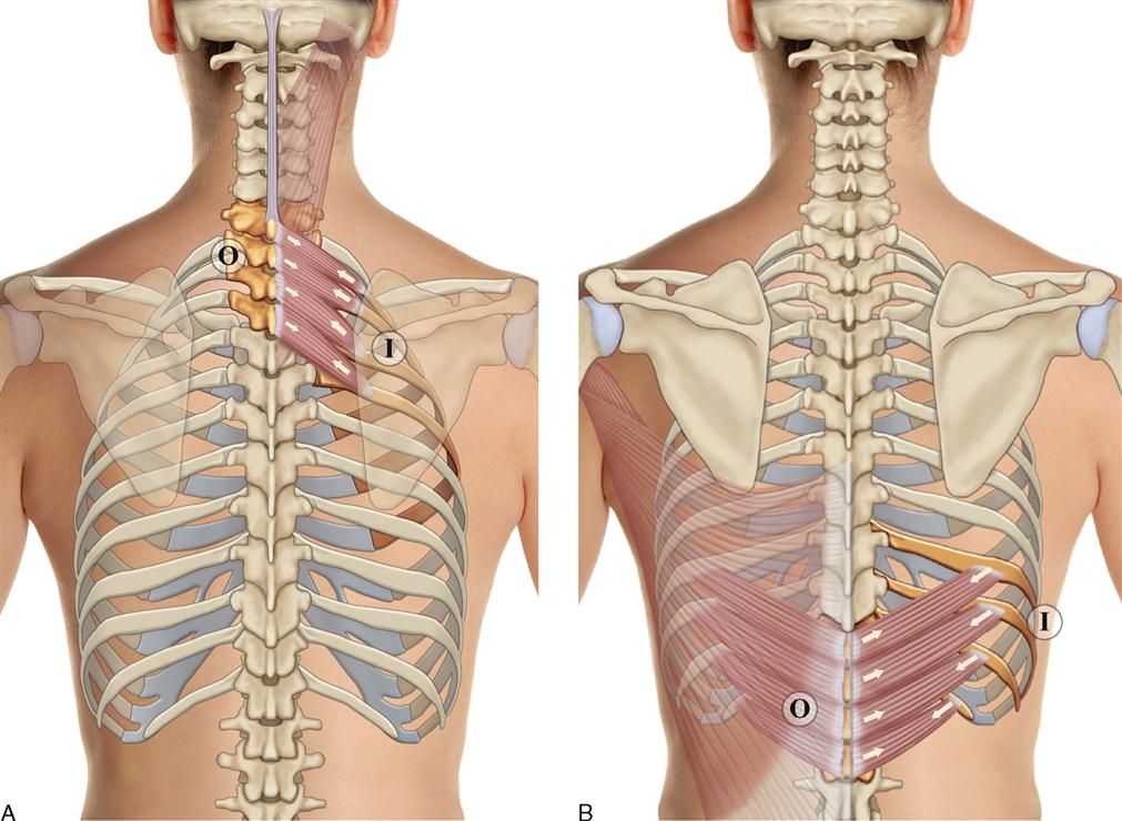
Each muscle elevates the rib immediately beneath the one it is attached to (can move individually or collectively to move the ribcage function:
Human skeleton system rib cage posterior view anatomy. Perspective front and back views. Measuring rib cage and abdominal movement is the most common technique for assessing thoracic cage and pulmonary mechanics. The teres minor is a narrow, elongated muscle of the rotator cuff. The thoracic cage (rib cage) is the skeletal framework of the thoracic wall, which encloses the thoracic cavity. There are many possible causes of rib cage pain. The rib cage, shaped in a mild cone shape and more flexible than most bone sets, is made up of varying elements the twelve pairs of ribs, which are embedded within the walls of the muscular structures, attach in the posterior to a thoracic vertebra. Related online courses on physioplus. It may occur after an obvious injury or without rib cage pain can be caused by a variety of things, ranging from pulled muscles to a rib fracture. Intercostal muscles are a group of muscles which exist in the intercostal space and help create from lateral border of sternum to the angle of rib (posteriorly it continues as posterior. The serratus posterior inferior and superior. It is supplied with blood by the lowest posterior intercostal artery, the subcostal artery articular cartilage of left superior articular facet of sacrum. On a muscular person when the muscles stretch, we see some of the.
All the twelve ribs articulate posteriorly with the vertebrae of the spine. Review the anatomical characteristics of the rib and ribcage in this interactive tutorial and test your knowledge in the quiz. Human rib cage anatomy model. A doctor will diagnose the underlying cause by a physical examination and imaging scans. Rib cage pain may be sharp, dull, or achy and felt at or below the chest or above the navel on either side.
Ch. 2 Part 4 - Communicative Disorders And Science 3100 ... from classconnection.s3.amazonaws.com Stretch those often forgotten rib muscles to relieve back pain and improve your posture. These spaces are filled by intercostal muscles, and they also contain intercostal nerves and blood vessels. This is an online quiz called rib cage muscles. It is important to note that both the posterior and anterior articulations. Saved by abbie betinis, composer. Rib cage pain may be sharp, dull, or achy and felt at or below the chest or above the navel on either side. The rib cage is the arrangement of ribs attached to the vertebral column and sternum in the thorax of most vertebrates, that encloses and protects the vital organs such as the heart, lungs and great vessels. So what parts of the rib cage show up on the surface?
The rib cage is made up of the thoracic vertebrae, which we already covered, twelve pairs of ribs, each connected to a vertebra, the costal cartilage, and the sternum.
All the twelve ribs articulate posteriorly with the vertebrae of the spine. The posterior or back muscles perform a wide range of functions, including movement of the shoulder, head, and neck and assisting in respiration, posture. Central slip and mallet finger management online course: Related online courses on physioplus. We're going to look at a pair of them that do just that: So what parts of the rib cage show up on the surface? Rib cage pain may be sharp, dull, or achy and felt at or below the chest or above the navel on either side. 308), is an osseocartilaginous cage which contains and protects the principal organs of respiration and circulation. These rib muscles automatically get worked when you do bench presses, push ups and dips, but a few bonus exercises can help you really zero in for a more chiseled torso. The thoracic cage (rib cage) forms the thorax (chest) portion of the body. Both the rib cage and the pelvis are important units of body structure; Structure of a typical rib: Review the anatomical characteristics of the rib and ribcage in this interactive tutorial and test your knowledge in the quiz.
So what parts of the rib cage show up on the surface? Rib cage pain may be sharp, dull, or achy and felt at or below the chest or above the navel on either side. Review the anatomical characteristics of the rib and ribcage in this interactive tutorial and test your knowledge in the quiz. Connective tissue of rib cage. Perspective front and back views.
Spine and Ribcage, Posterior View - Medical Illustration ... from www.doereport.com On a muscular person when the muscles stretch, we see some of the. The skeleton of the thorax, or chest (fig. We're going to look at a pair of them that do just that: These spaces are filled by intercostal muscles, and they also contain intercostal nerves and blood vessels. The serratus posterior inferior and superior. This is an online quiz called rib cage muscles. The posterior or back muscles perform a wide range of functions, including movement of the shoulder, head, and neck and assisting in respiration, posture. Related online courses on physioplus.
Muscles of the spine and rib cage | musculoskeletal key.
Central slip and mallet finger management online course: Compresses the lower portion of the rib cage (can elevate lower ribs if humerus is fixed thus, can generate active force of inspiration and. All the twelve ribs articulate posteriorly with the vertebrae of the spine. A doctor will diagnose the underlying cause by a physical examination and imaging scans. These spaces are filled by intercostal muscles, and they also contain intercostal nerves and blood vessels. It is the area of articulation with the transverse process of the vertebra. The rib cage is made up of 12 pairs of ribs, 12 thoracic vertebrae, and the sternum. The rib cage, shaped in a mild cone shape and more flexible than most bone sets, is made up of varying elements the twelve pairs of ribs, which are embedded within the walls of the muscular structures, attach in the posterior to a thoracic vertebra. For this purpose isolated strips of rib cage elevator muscle of variations in the musculoskeletal system exist in different classes of animal. Each muscle elevates the rib immediately beneath the one it is attached to (can move individually or collectively to move the ribcage function: Perform dumbbell pullovers to work the muscles along your rib cage. Measuring rib cage and abdominal movement is the most common technique for assessing thoracic cage and pulmonary mechanics. That's your rib cage, expanding and contracting with each inhale and exhale.
Related online courses on physioplus rib cage muscles. Constant sitting (and especially straining your neck to look down while sitting) causes tightness in the front of the ribs and puts stress on the back.


0 Comments:
Posting Komentar